IRAMED is a €550.000 Prometeo project funded by Generalitat Valenciana that focuses its studies on optimizing exposure to ionizing radiation in treatments and radiodiagnosis, so that the doses received by patients are reduced as much as possible without affecting the quality and accuracy of treatment or diagnosis.
KEY CHALLENGES
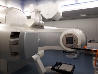
The harmful effect that ionizing radiation has on people’s health is well known. Despite this, in the medical field, the diagnosis of diseases, as well as certain medical treatments (especially cancer) justifies its use in patients.
The great challenge of the IRAMED project is to reduce the doses that patients receive when a clinical diagnosis is made through Computed Tomography or PET-CT applications or when a radiotherapy or protontherapy treatment is performed on the patient.
THE SOLUTION
To address these challenges, it will be necessary to develop new numerical methods and simulations of particle transport according to the case study, mainly using Monte Carlo codes for the simulation of particle transport.
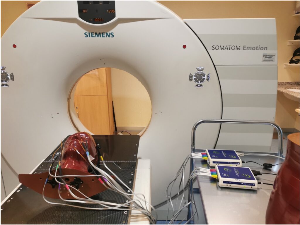
To reduce the dose in CT and PET-CT, the Monte Carlo numerical simulation codes (PenRed, GEANT, PENELOPE, GATE, GAMOS and MCNP) and the databases provided on the Cancer Imaging Archive website will be used. From sinograms and reconstructed images, filters based on Artificial Intelligence will be developed to reduce artifacts, noise and improve image quality.
Its success will guarantee new systems for diagnosing cancer, mental illnesses and other pathologies in the future, optimizing irradiation doses for patients.
On the other hand, the precision in radiotherapy treatments will be improved, as well as the reduction of unwanted secondary doses to patients and staff generated by photoneutrons induced in high energy radiotherapy treatments (Linear Accelerator -Linac-) and protontherapy will be studied.
OBJECTIVES
The great challenge of the IRAMED project is to reduce the doses that patients receive when a clinical diagnosis is made through Computed Tomography or PET-CT applications or when a radiotherapy or protontherapy treatment is performed on the patient.
The specific objectives set out in the IRAMED project are:
-Reduction of radiation dose in radiodiagnosis, specifically in Computed Tomography.
-Reduction of radiation dose in Nuclear Medicine, particularly in PET-CT technology.
– Minimization of radiation dose in radiotherapy and protontherapy treatments.

METHODOLOGY
O1. CT DOSE REDUCTION
CT can be used for radiodiagnosis and for planning a radiotherapy treatment. Therefore, this objective proposes, on the one hand, the estimation and reduction of radiation dose through simulation using the PenRed Monte Carlo code and, on the other hand, the application of medical image reconstruction techniques with few projections using high-performance tools (HPC), especially 3D TC and Cone-Beam applications. In addition, new image filtering techniques will be developed based on Artificial Intelligence techniques.


O2. PET-CT DOSE REDUCTION
The proposed line of work consists, on the one hand, of optimizing the methods for calculating doses in the PET technique using the Monte Carlo method and, on the other hand, developing a methodology to compare the real images obtained by PET (in tests clinics) with images reconstructed from Monte Carlo simulation. The confluence of the objectives will allow dose maps to be associated with each PET image and, consequently, it will be possible to provide information on possible dose reduction.


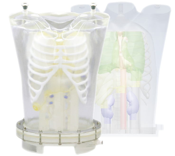
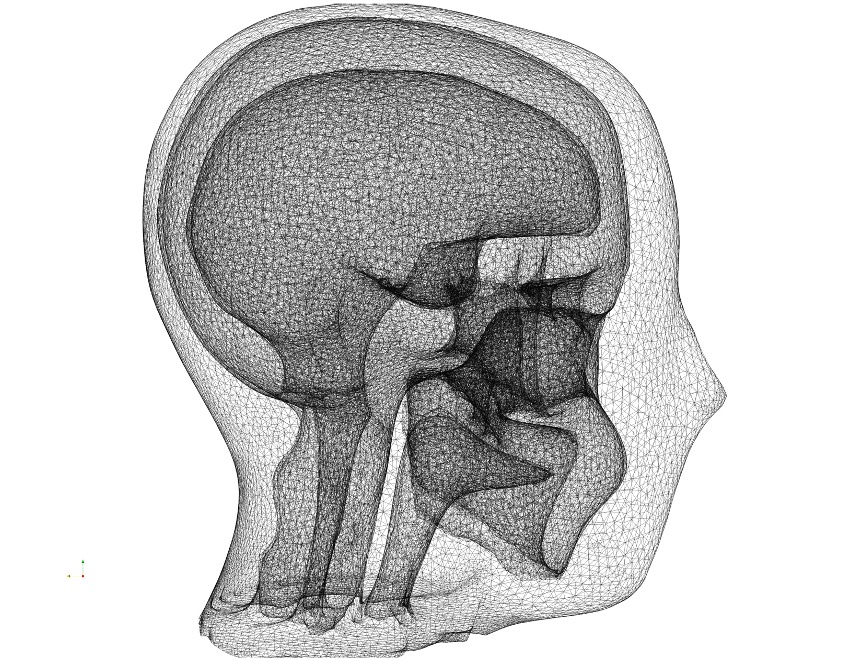
The line of research includes the verification of models of analytical and anthropomorphic phantoms with the GATE software, based on the Monte Carlo method. This simulation tool together with the image reconstruction software allows obtaining radiodiagnostic images.
The line includes modeling PET scanner prototypes with ring spacing to explore the potential of reduced-cost scanners.
Monte Carlo simulation allows developing proofs of concept to evaluate the potential of various new PET designs.


O3. RADIOTHERAPY AND PROTONTHERAPY DOSE REDUCTION
The proposed line of work consists, first of all, in obtaining the response function of the measurement equipment, Bonner Sphere Spectrometer (BSS). Experimental measurements will be carried out in the different spaces where the radiotherapy and protontherapy treatment equipment is located. The energy spectrum will be obtained from the measurements made using the response function matrix of the Bonner sphere system. Once the energy spectrum is known, the dose absorbed by patients at the points considered in the room can be estimated, thus allowing improvement in cancer treatments and reducing secondary cancers due to secondary radiation.


04. COMMUNICATION, DISSEMINATION AND EXPLOITATION OF RESULTS
A fundamental objective of the project is that the studies and results obtained during the project are in accordance with the current needs and interests of society. The focus of the project will have a scientific/technological impact and it is of utmost importance to carry out an exhaustive communication, dissemination and exploitation campaign of the results derived from the project. We will collaborate with hospitals in the Valencian Community and companies in the physical-medical engineering sector to further disseminate the results of the project and we will communicate with different target audiences.

IMPACT
The hypothesis on which this project is based is quite simple: the estimation of the doses of organs that patients receive during diagnosis Computerized Tomographies is relevant information.
Despite the efforts of the radiology community recommending appropriate radiation dose estimation procedures consistent with the ALARA (As Low As Reasonably Achievable) criteria, studies have been published showing that there is great variability in the dose imparted, within the same institution and between different institutions, in the diagnostic dose CT image.
The direct benefit of this project is very important, since the proposed tool to determine the given dose will be fast and accurate, with the possibility of integrating it into the patient’s dosimetry history, which in the long term will improve public health.
In addition, tools will be developed that allow the reconstruction of images with applications to 3D CT and Cone-Beam with few projections, thus reducing the time of exposure to radiation for patients. Additionally, AI methods will be developed to remove noise from CT images to improve image quality.
On the other hand, high-energy radiotherapy and protontherapy are associated with fast and thermal photoneutrons. In fact, the IAEA and NCRP have recommended protocols for calculating adequate shielding against contaminating neutrons. The study of these neutrons will make it possible to improve the designs of the facilities, but above all it will allow the improvement of the radiological protection of the patients and staff of these facilities.
