O1. TC DOSE REDUCTION
Computed tomography (CT) is a diagnostic imaging technique that combines multiple X-ray images taken from different angles to create detailed cross-sections of the human body. These images allow doctors to accurately visualize the body’s internal structures, including organs, soft tissues, bones, and blood vessels.
CT is widely used in the diagnosis and monitoring of a variety of medical conditions, such as traumatic injuries, cardiovascular diseases, cancer, neurological disorders, and more. It provides detailed, accurate images that can help doctors detect and diagnose diseases, plan treatments, and evaluate the effectiveness of medical interventions.
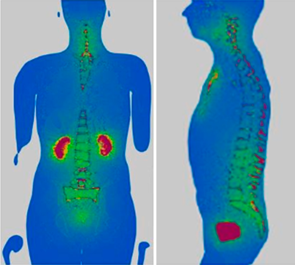
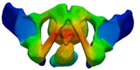
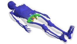
This diagnostic technique is an invaluable tool for studying internal structures of the human body and diagnosing a wide range of medical conditions. However, it is crucial to address the risks associated with exposure to ionizing radiation and look for ways to optimize exposure to reduce the dose received by patients.
In this context, IRAMED project researchers are working on the development of image reconstruction techniques with few projections. New imaging techniques are being explored and developed that can provide high-quality images with lower radiation doses. This includes the use of low-projection image reconstruction algorithms and AI filtering techniques to improve image quality while reducing radiation dose.
On the other hand, they are applying Monte Carlo techniques, with the Monte Carlo Penred code, to calculate doses and thus develop a code to plan radiotherapy treatments.

1. Radiation dose estimation by Monte Carlo methods
CT can be used for radiodiagnosis or treatment planning. Since ionizing radiation became a therapeutic application, the calculation of dose distribution has been a problem due to the complexity of treatment planning. When dose gradients or complex distributions appear in a treatment (in IMRT, IGRT or in radiosurgery, for example) or when the tissue to be irradiated is very heterogeneous, the algorithms used in current systems can give values outside the allowed margins for the health.
It has been shown that Monte Carlo methods and those based on ETB applied to the transport of ionizing radiation are the best methods to calculate doses in any type of medium and complex geometries considering the physics involved in the problem.
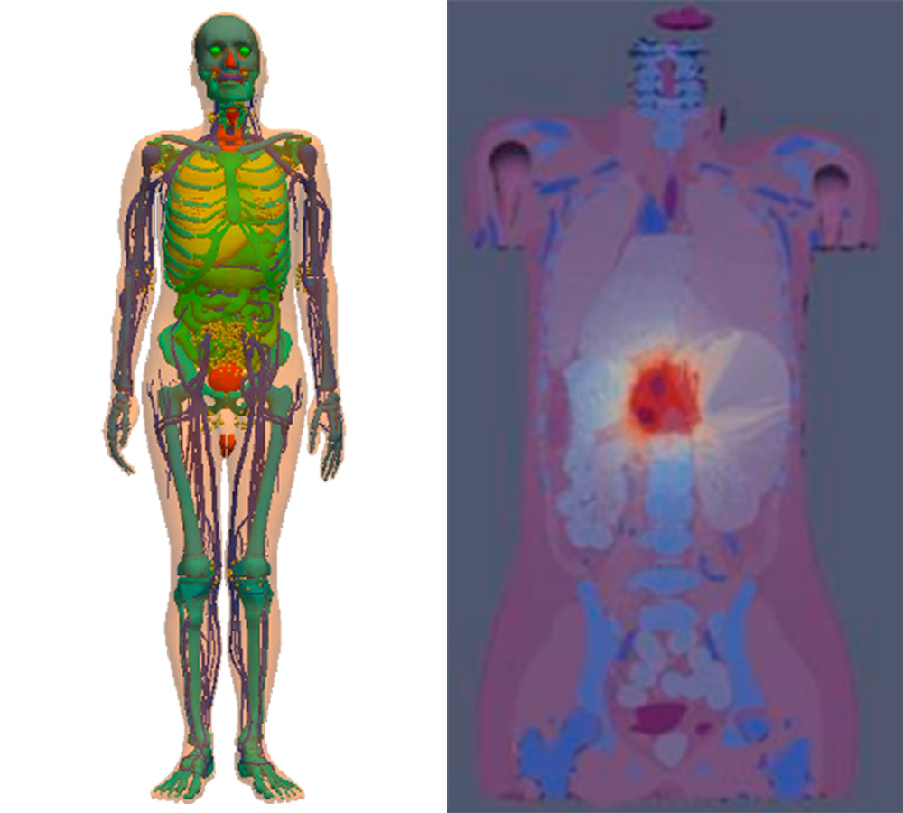
From the CT scans performed in radiotherapy for planning, simulation and verification, it is possible to reduce the dose. Along these lines, the Monte Carlo PenRed code can simulate treatment planning for patients, also reducing doses in Teletherapy and Brachytherapy, and to do so, the code will be adapted and verified.
2. Image reconstruction techniques with low-projections
The Computed Tomography (CT/CT) medical test is currently essential in clinical practice for the diagnosis and monitoring of multiple diseases and injuries, being one of the most important medical imaging tests due to the large amount of information it is capable of providing. However, unlike other imaging methods that are harmless, the CT test uses x-rays, which are ionizing, so they pose a risk to patients.
This is why it is necessary to develop methods that allow reducing the radiation dose to which patients undergoing a study are exposed, without compromising image quality, since otherwise these people would be exposed to risk without the benefit (a quality diagnosis) is guaranteed.
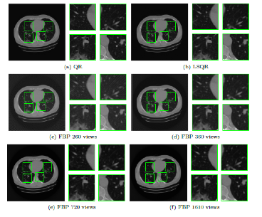
During the development of this line, CT image reconstruction methods are being investigated that are based on reducing the number of projections used, with the aim of reducing the exposure time to X-rays. This dose reduction strategy is in the development phase research, unlike others that are implemented in clinical practice and have already been developed by the scanner manufacturers themselves. It will be applied to 3D CT and Cone-beam and high-performance computational techniques will be used to reduce computing times.
3. Artificial Intelligence applications
The most important challenge consists of creating a filter based on Artificial Intelligence (Deep Learning Neural Network). The input would be the sinograms or the reconstructed signal with low dose, the reference the reconstructed signal with high dose, and the solution of the filter parameters that through neural networks make the reconstructed output file (CT image) optimally approximate the reference file.
